
Predicting Prostate Cancer
Machines that think for themselves – beyond being mere tools for humans – have fascinated writers and filmmakers for decades. Depictions of "beings" possessing artificial intelligence have ranged widely from the compassionate protagonist, David, in Steven Spielberg's film A.I. to the dangerous computer villain, HAL, in Stanley Kubrick's film 2001: A Space Odyssey.
While the promise and peril of this technology continue to be debated by scholars and authors of fiction, applications of artificial intelligence quietly abound in daily life, enabling services such as bank fraud prevention programs, facial and voice recognition, and research into self-driving automobiles. Biomedical scientists are quickly embracing the technology as a way to improve analysis of massive amounts of data, such as screening patients for eligibility in clinical trials by filtering numerous eligibility criteria simultaneously. At the Medical College of Wisconsin, scientists are applying the technology to enhance what future radiologists will see when looking at MRI scans of the prostate, brain and potentially other organs.
Peter LaViolette, PhD '11, MS, MCW assistant professor of radiology and biomedical engineering, began his scientific career as a high-energy nuclear physics researcher. After several years, however, he knew that he needed to make a change. "I loved doing research, but I was just too disconnected from relevant medical applications," he says. Dr. LaViolette, while working in imaging software development at Massachusetts General Hospital and considering moving back to his home state of Wisconsin, learned from a colleague about MCW's pioneering history in medical imaging and functional MRI. This prompted him to enroll at MCW, where he earned a doctoral degree in biophysics.
"My imaging research at MCW began in brain cancer, as I was interested in comparing physical tissue to MRI scans to improve what we can extract from the MRI data. No one had programmed software to conduct this comparison, so we built it ourselves," explains Dr. LaViolette. While doing so, he realized he could further analyze the data generated by applying machine learning.
Machine learning is a radically different approach to interfacing with a computer than traditional programming. Rather than discretely telling the computer every parameter of a problem with handwritten code, machine learning developers and scientists apply artificial intelligence to set up frameworks whereby computers teach themselves. In games like chess, for example, developers prepare a handful of algorithms that enable a computer to read and evaluate the moves and outcomes of a massive number of matches. By learning how moves and positioning affect wins and losses, machine learning programs have demonstrated the ability to routinely predict the best possible move in each scenario which has enabled machines to compete with elite chess players who have honed their skills over many years.
For Dr. LaViolette, a similar approach allows his lab to teach a computer how to predict the location and severity of prostate tumors from the data in MRI scans.
"While Dr. LaViolette continues to study brain imaging, we realized that the prostate was an excellent model for machine learning research because it is a less complex organ, and we can draw on better definitions of tumor severity," remarks Sean McGarry, an MCW graduate student working in the LaViolette lab.
Kenneth A. Iczkowski, MD, MCW, professor of pathology and urology who collaborates with Dr. LaViolette, notes, "As a pathologist interested in how tumor growth patterns affect treatment and clinical outcomes, I jumped at the chance to work together. Others have tried to correlate the cancer patterns I analyze on glass slides with MRI scans, but never as precisely as this."
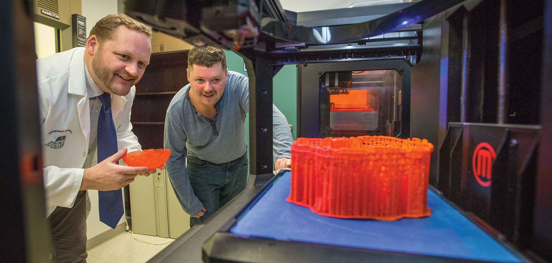
Patients whose cancerous prostates are being removed and who agree to participate have an MRI scan taken before surgery. Afterwards, Dr. LaViolette uses dimensions calculated from the MRI scan to print a custom 3-D mold of each prostate. This mold allows the prostate to be sliced and processed into glass slides that precisely match the MRI scan. Dr. Iczkowski then analyzes the tissue for tumor severity (grade), growth pattern and location, and sends annotated images to Dr. LaViolette. Finally, Dr. LaViolette and McGarry feed the annotated pathology and the MRI data into a machine learning program. By comparing the images, the software learns what features in the MRI are predictive of tumors of varying severity or growth pattern.
The long-term goal is to improve the staging and monitoring of patients who have prostate cancer but may not need surgery. It also may allow for more targeted biopsies and radiation therapy, which would improve the accuracy of diagnosis and reduce side effects from damage to healthy tissue. This aligns well with advances in the overall field of prostate cancer treatment.
"More and more, active surveillance and watchful waiting are being seen as reasonable approaches. Not every prostate cancer case is aggressive and needs immediate surgery or radiation. But we need more information to get better at monitoring patients and staging their treatments," says Dr. LaViolette.
Within the next five years, Dr. LaViolette believes he and colleagues from radiology, radiation oncology, urology and pathology will be ready for a clinical trial to improve the targeting and dosing of radiation therapy for prostate cancer. Until then, he, McGarry and Dr. Iczkowski will continue to fine-tune their machine learning algorithms and improve their power to predict tumor characteristics by interpreting MRI scans.
"I really enjoy this lab because Dr. LaViolette staunchly supports our freedom of curiosity. That ability to explore is what will allow us to continue advancing this science," comments McGarry. Dr. Iczkowski adds, "As we improve, the potential is very promising to apply this approach to other solid organ tumors."
Recommended